
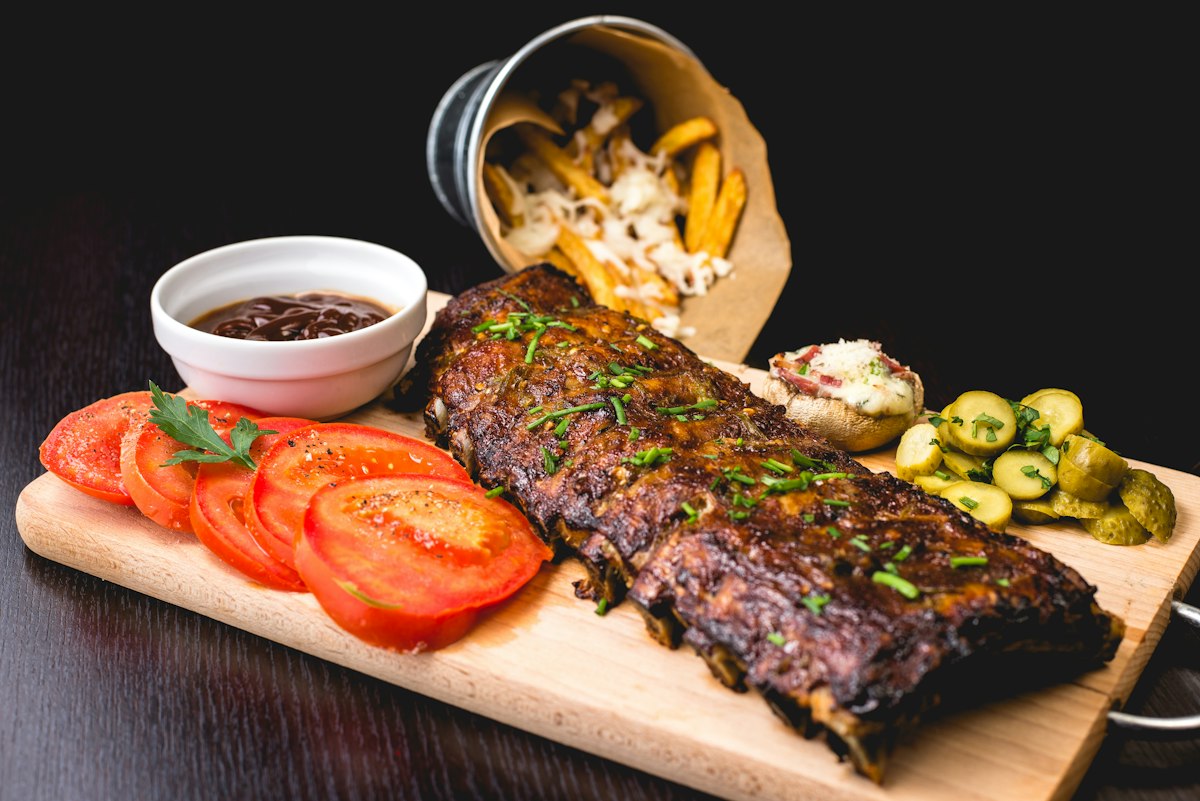
Dietary Advanced Glycation End-Products (dAGEs)—a heterogeneous group of non-enzymatic adducts formed when reducing sugars react with free amino groups on proteins, lipids, and nucleic acids—represent a paramount pro-inflammatory dietary hazard. Accelerated exponentially by high-temperature, low-moisture culinary techniques (dry roasting, deep-frying, grilling, and broiling), dAGEs escape complete digestive proteolysis, undergo systemic intestinal absorption, and bind to vascular receptors, promoting accelerated atherosclerosis, endothelial dysfunction, and end-organ microvascular cross-linking.
1. Introduction: The Classical Maillard Cascade and Major dAGE Molecular Moieties
The genesis of dAGEs initiates via the nucleophilic condensation of reactive carbonyls (glucose, fructose, ribose) with the epsilon-amino group of lysine or the guanidino group of arginine residues. This generates a reversible Schiff base that spontaneously isomerizes via Amadori rearrangement into stable 1-amino-1-deoxy-2-ketoses (such as fructoselysine).
Under the catalytic influence of transition metals (Fe²⁺/Cu²⁺) and atmospheric oxygen (Hodge scheme oxidation), Amadori intermediates undergo oxidative fragmentation (Namiki and Hodge pathways) into highly reactive alpha-dicarbonyl intermediates: methylglyoxal (MGO), glyoxal (GO), and 3-deoxyglucosone (3-DG). These potent electrophiles react rapidly with protein amino residues to generate irreversible dAGE structures:
N-epsilon-(carboxymethyl)lysine (CML): The dominant analytical surrogate biomarker for dietary glycation, formed via oxidative cleavage of fructoselysine or reaction of glyoxal with lysine.
Methylglyoxal-Derived Hydroimidazolone 1 (MG-H1): The primary quantitative dAGE adduct derived from the reaction of methylglyoxal with arginine guanidino groups.
Glucosepane: An irreversible lysine-arginine cross-linking adduct responsible for the mechanical stiffening of human extracellular matrix proteins.
2. Technical Analysis: The RAGE Inflammatory Axis and Intercellular Collagen Stiffening
Following the ingestion of high-dAGE foods, approximately 10% to 30% of dietary glycation adducts are absorbed across the intestinal epithelium via paracellular diffusion (for free low-molecular-weight adducts) or via peptide transporter 1 (PEPT1, for di- and tripeptide-bound AGEs). Once in systemic circulation, dAGEs engage two pathological cascades:
RAGE Axis Signal Transduction: Circulating dAGEs bind to the immunoglobulin-superfamily Receptor for Advanced Glycation Endproducts (RAGE) expressed on vascular endothelial cells and macrophages. Ligand-RAGE engagement stimulates membrane-bound NADPH oxidase (NOX1 and NOX4), generating massive bursts of intracellular superoxide (O₂•⁻) and hydrogen peroxide (H₂O₂). This oxidative storm phosphorylates the I-kappa-B kinase (IKK) complex, releasing Nuclear Factor kappa B (NF-kappa-B) to translocate into the nucleus, driving sustained transcription of pro-inflammatory cytokines (IL-6, TNF-alpha) and vascular cell adhesion molecule 1 (VCAM-1).
Vascular Extracellular Matrix Cross-Linking: Independent of receptor signaling, reactive dAGE precursors form permanent covalent intermolecular cross-links (glucosepane and pentosidine) between adjacent tropocollagen triple helices in arterial elastic laminae. This cross-linking eliminates matrix elastance, escalating arterial pulse-wave velocity (PWV) and driving systolic hypertension:
| Dietary dAGE Biomarker | Precursor Intermediates | Typical High-Heat Food Concentration | RAGE Binding Affinity K_d (nM) | Pathological Vascular Consequence |
|---|---|---|---|---|
| N-epsilon-(carboxymethyl)lysine (CML) | Fructoselysine / Glyoxal + Lysine | 5000 – 12000 μg/100g (broiled meat, bacon) | 140 – 220 nM | Endothelial NO uncoupling (eNOS down-regulation) |
| MG-H1 (Hydroimidazolone) | Methylglyoxal + Arginine | 8000 – 18000 μg/100g (fried snacks, cheese) | 85 – 120 nM | Macrophage foam cell formation via CD36 |
| Glucosepane | D-Glucose + Lysine + Arginine | Progressive tissue accumulation | Structural cross-link | Loss of vascular arterial compliance (+28% PWV) |
| Pentosidine | Ribose + Lysine + Arginine | 300 – 800 μg/100g (roasted coffee, crusts) | Fluorescent cross-link | Diabetic nephropathy & glomerular stiffening |
3. Conclusion & Industrial Implications: Wet-Culinary Modifications and Molecular Trapping Agents
Mitigating dietary dAGE accumulation in culinary processing relies on thermodynamic manipulation: cooking with moist heat (poaching, steaming, stewing, sous-vide at T < 100°C) rather than dry heat (roasting, grilling at T > 180°C) reduces dAGE formation by over 80%. Furthermore, pre-marinating proteins in acidic solutions (lemon juice, vinegar, pH < 4.0) protonates lysine amino groups, competitively inhibiting initial Schiff base condensation. Supplementation with endogenous dicarbonyl scavengers—specifically the dipeptide carnosine (beta-alanyl-L-histidine) and pyridoxamine (vitamin B6)—traps alpha-dicarbonyls into harmless adducts, preserving vascular compliance and metabolic longevity.